

Peroneal Tendons
What are the Peroneal Tendons?
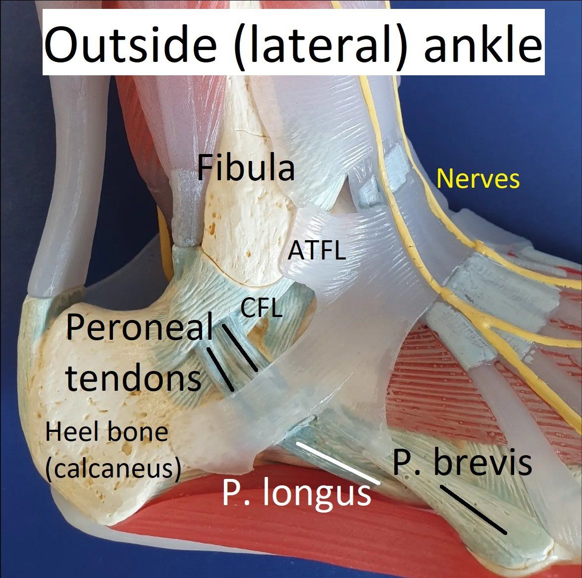
The peroneal tendons run down the outside of the leg and ankle to connect with the foot.
There are two important tendons:
- peroneus brevis
- peroneus longus.
They both act to keep the ankle and foot stable on uneven ground. This helps protects the ankle ligaments from injury.
See image
and Ankle - Anatomy and Imaging.
What can go wrong with the Peroneal Tendons?
The peroneal tendons help maintain ankle stability
and balance on uneven ground. When there is ankle instability
or the foot has a high arch, they can become overloaded and damaged. This can cause:
- peroneal tendinopathy ("wear and tear")
- split tears
- inflammation.
Occasionally, an ankle injury can make these tendons unstable.
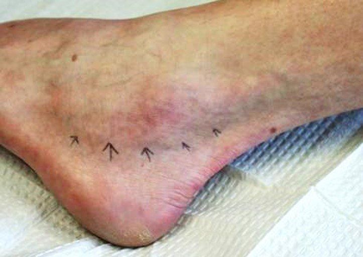
Peroneal Tendinopathy and Split Tears
When the peroneal tendons are chronically overloaded, they start to fray and thicken. This is called tendinopathy. Symptoms are often mild.
If this causes inflammation or the tendon splits, significant pain and swelling develop behind the fibula and down the outer foot (see image).
Rarely, a peroneal tendon can rupture. This is usually after months of inflammation and pain.
Peroneal Tendon Instability
The peroneal tendons are held in place behind the fibula by a strong ligament (superior peroneal retinaculum). This ligament can be torn allowing the tendons to slip forward (dislocate) over the fibula.
People with this injury have painful swelling and clicking
on the outside of the ankle. The ankle can feel unstable.
Interesting fact: 5% of people are born with an extra (accessory) peroneal tendon called peroneus quartus that can make the tendons click.
Conditions associated with Peroneal Tendon Problems
Any condition that makes the peroneal tendons work harder increases the chance of them developing a problem.
Some of these are:
- ankle instability (see image below)
- high arched (cavo-varus) feet (see image below)
- Achilles and calf tightness
- generalised ligament laxity
- previous calcaneus fracture.
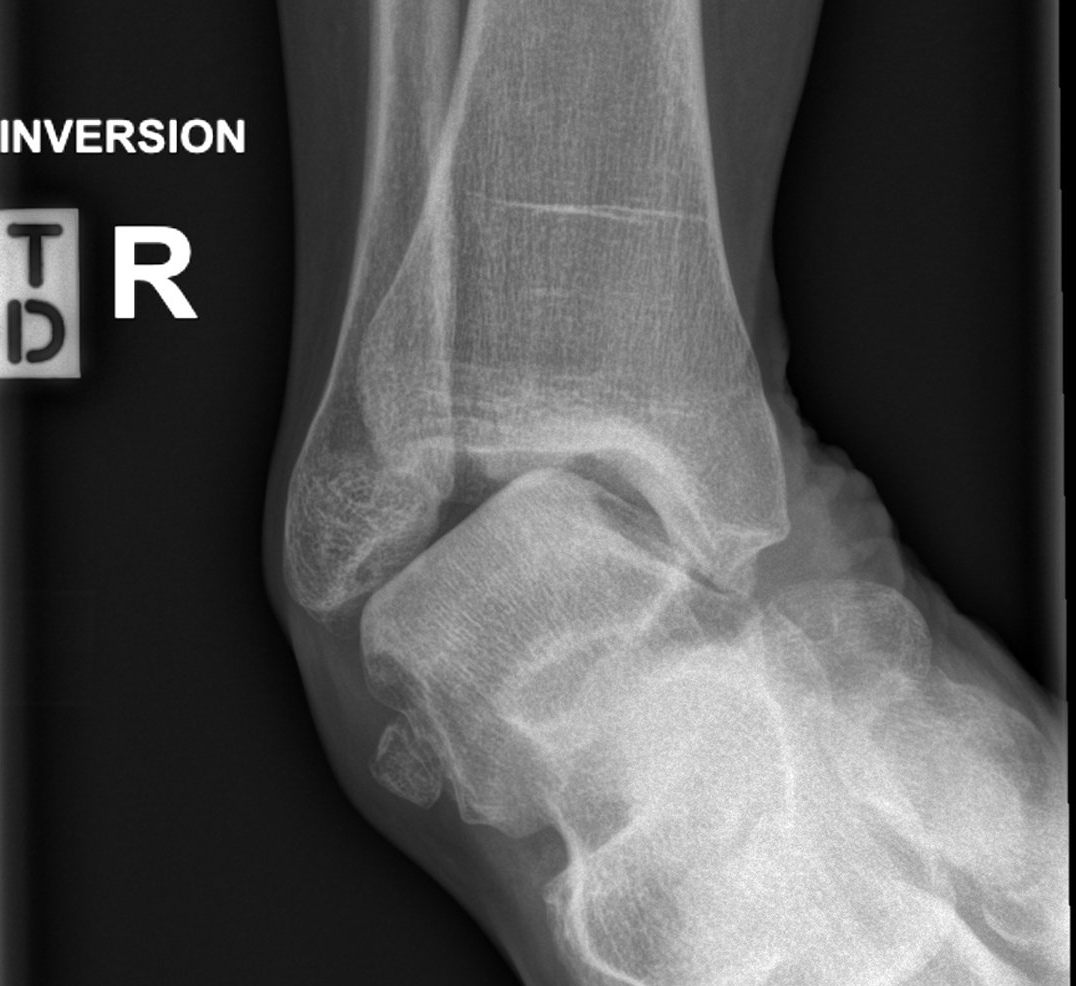
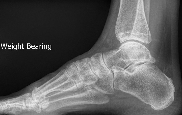
How are Peroneal Tendon Problems Diagnosed?
The diagnosis is usually clear after taking a complete history and performing a physical examination. It is critical that ankle stability and arch height are assessed. Peroneal tendon instability can only be diagnosed by careful examination.
Investigations commonly arranged are:
- weight-bearing X-rays of the ankle and foot
- ultrasound and/or MRI scan of the ankle and peroneal tendons.
How is Peroneal Tendinopathy Treated?
Most people improve with non-surgical treatment unless a large split tear is present.
Non-surgical treatment
Non-surgical treatment involves:
- reduced activity on uneven ground
- anti-inflammatory medications
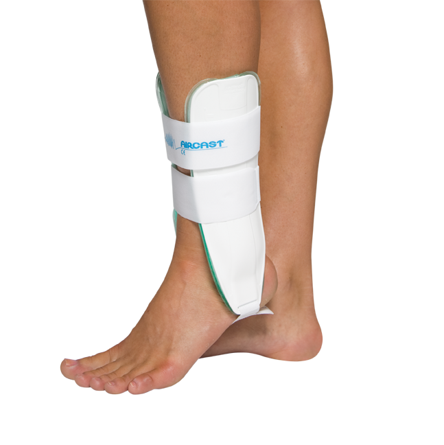
- ankle braces and supportive shoes
- physiotherapy
- podiatry and insoles for people with high arches
- cortisone injections.


Surgery for peroneal tendinopathy and split tears
If symptoms are not improving after several months of non-surgical treatment or a large split tear is present, surgery is often recommended.
The aim of surgery is to assist tendon healing and correct any associated condition. This usually involves:
- ankle arthroscopy
- peroneal tendon exploration with repair or reconstruction.
Other possible procedures include:
- ankle stabilisation
- 1st metatarsal dorsi-flexion osteotomy
- calcaneal minimally-invasive osteotomy.
Recovery usually requires a "moon-boot" for six weeks then physiotherapy. Full recovery takes at least six months. Recurrence can occur (15%).
How is Peroneal Tendon Instability Treated?
Pain-free peroneal tendon clicking does not need any treatment.
Significant peroneal tendon instability always requires surgery.
This involves open peroneal stabilisation with removal of any accessory muscle or tendon, and is 95% successful. It also requires six weeks in a "moon-boot".