
Calcaneus Fracture
What is a Calcaneal Fracture?
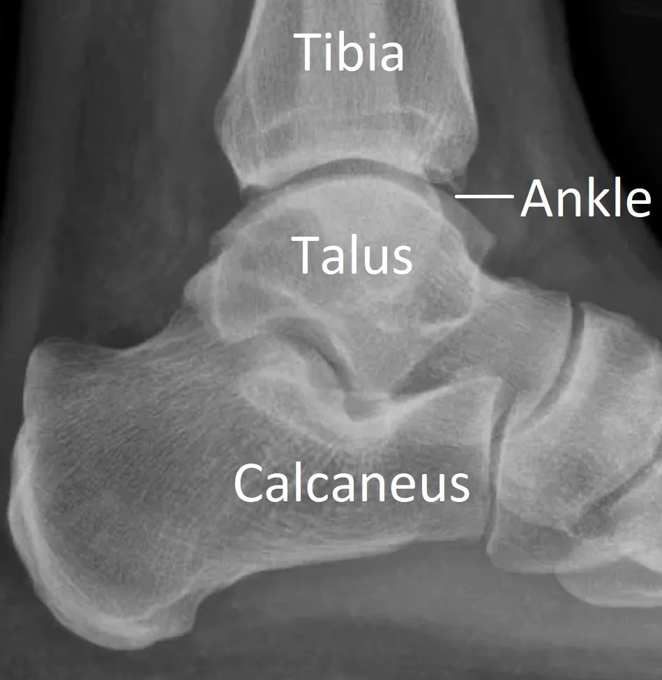
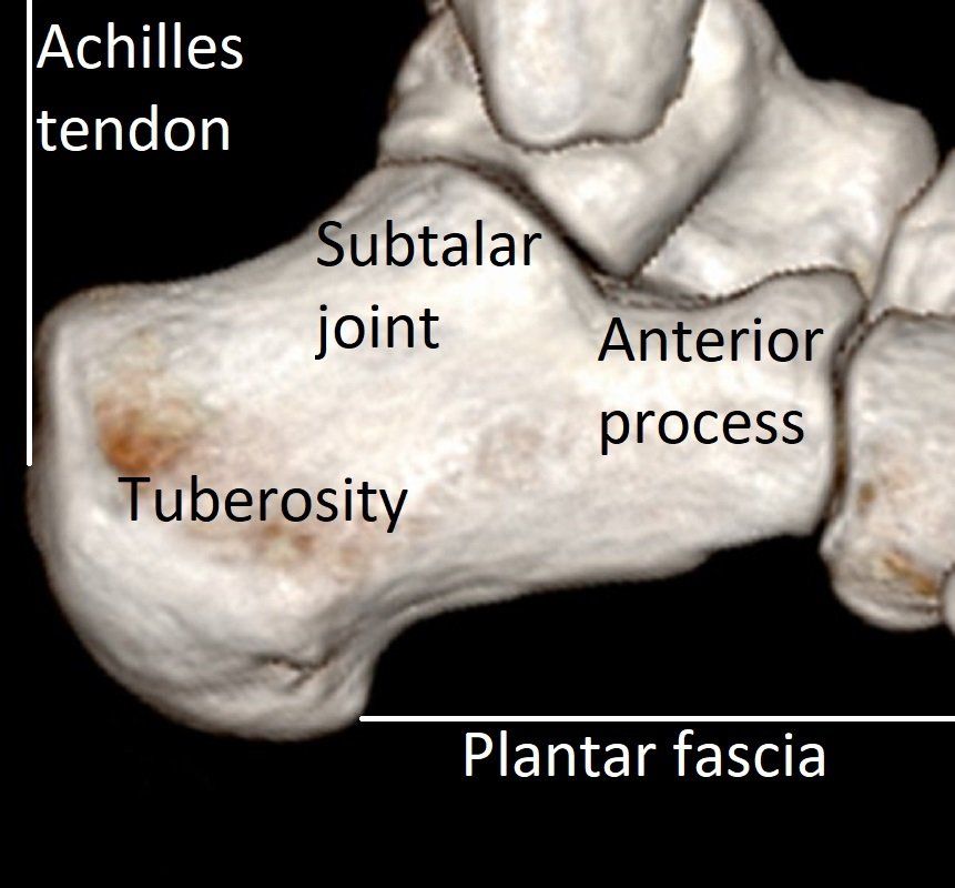
The calcaneus (or heel bone) is the largest bone in the foot. The calcaneus connects to other bones forming the subtalar and calcaneocuboid joints of the hindfoot. The Achilles tendon attaches to the back of the calcaneus.
A fracture is a break in a bone. Calcaneal fractures are uncommon but some are severe injuries. Healing can take many months and long-term complications occur if the joints are damaged or the bone flattened.
Causes of Calcaneal Fracture
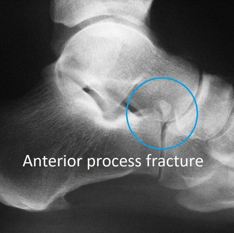
Minor
avulsion fractures of the anterior process occur with twisting injuries (see Why is my ankle sprain not getting better?).
Severe
fractures of the calcaneus are usually due to falling from a height, such as a ladder, and landing on the heel.
- This crushes the calcaneus making it spread out sideways and lose height.
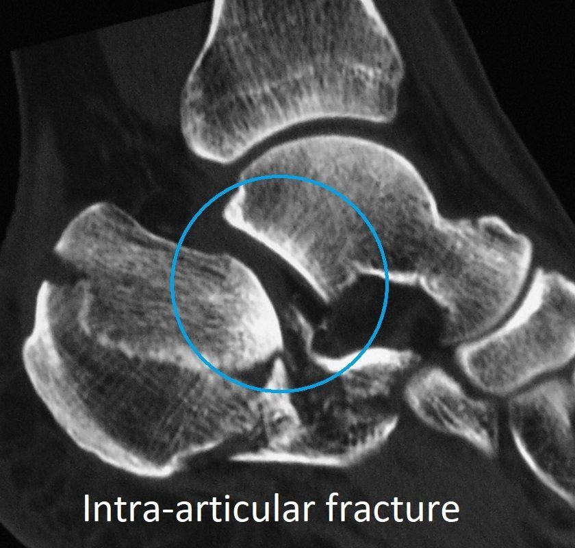
- Injury to the subtalar joint is common (intra-articular fracture).
Symptoms of Calcaneal Fracture
The common symptoms of calcaneal fractures are:
- pain in the heel
- swelling in the heel
- inability to walk or bear weight on the foot.
Diagnosis of Calcaneal Fracture
Foot injuries sustained in a significant fall or motor vehicle accident require the whole body be assessed for other injuries.
After taking a history and examining the ankle and foot, imaging is performed:
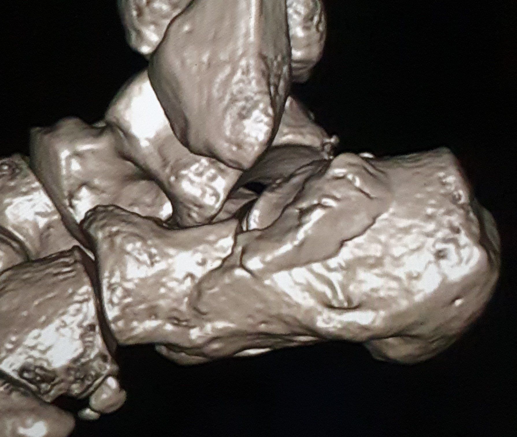
- X-rays
- CT scan.
These imaging investigations show which part of the calcaneus is broken, and how badly. This determines both the need for surgery and the risk of long-term complications. Severe injuries can lead to hindfoot osteo-arthritis.
Treatment of a Calcaneal Fracture
Calcaneal fractures are treated based on the type of fracture and extent of soft tissue damage.
The surgical treatment of calcaneal fractures continues to improve. Previous techniques involved lifting up large "flaps" of skin to get to the bone. This had a high complication rate that didn't justify surgery in most people. New techniques use smaller incisions and are safer.
Unfortunately, even a well fixed calcaneal fracture can develop subtalar osteo-arthritis. However, treating this by subtalar fusion is much easier than if the calcaneus is left flat and deformed.
Non-surgical treatment
- rest, ice, compression, and elevation (RICE) - is used for all calcaneal fractures.
- protection and crutches – using a "moon-boot" can be more comfortable in the first 3 weeks after injury but leads to hindfoot stiffness if used too long after this. Crutches, etc, are important for 10 weeks to avoid weight bearing on the foot until healing has occurred.
- movements - gentle ankle and foot movements need to start early to prevent stiffness. This is helped by being in a pool.
Surgical treatment
Surgery is only offered to selected people and calcaneal fractures. It is rarely appropriate in active smokers. Certain fracture patterns do better with surgery and others do not.
Surgical options are:
- mini-open reduction and internal fixation
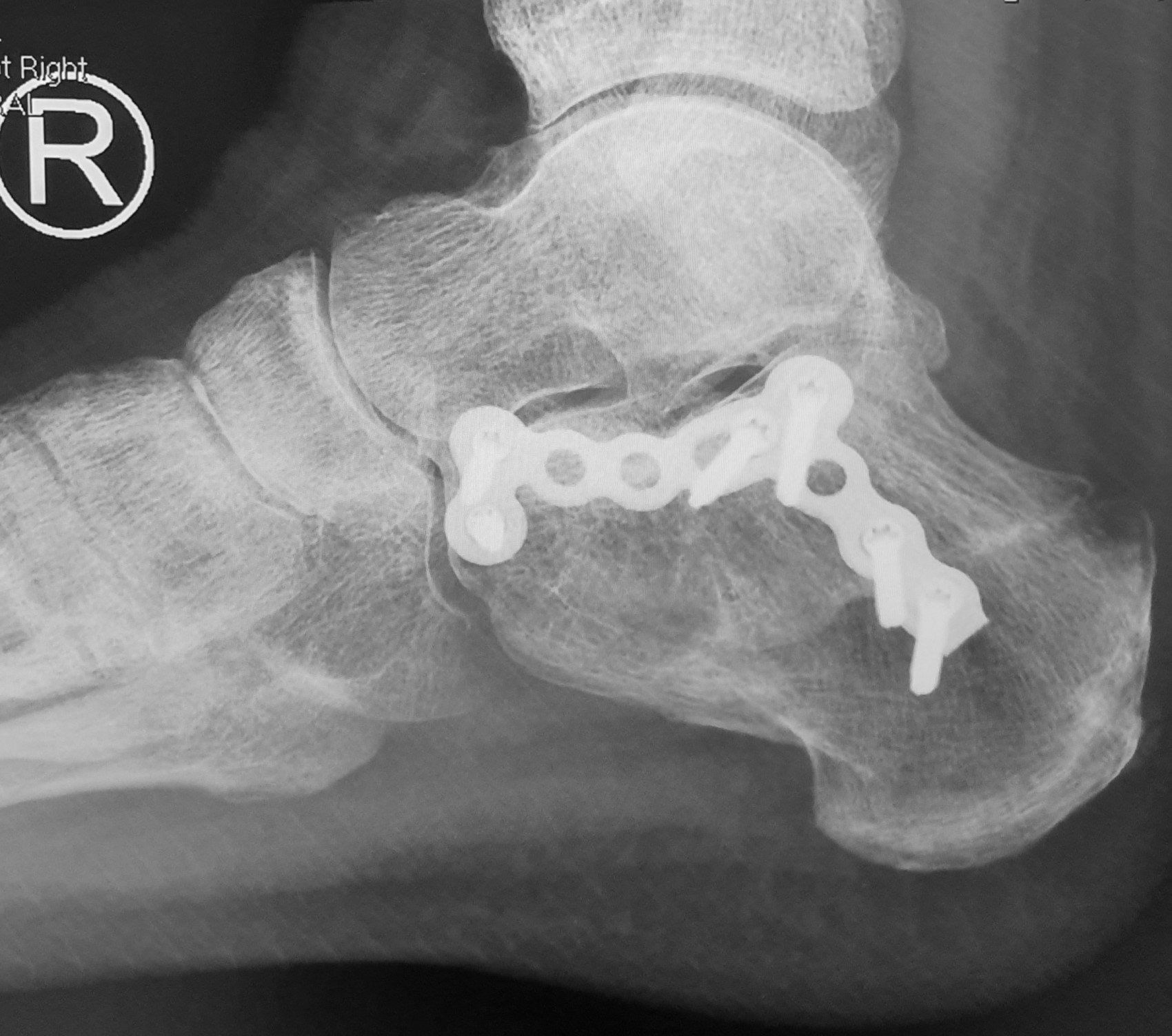
– this surgery involves putting the bone fragments back together with metal plates and screws. Dr Beamond has experience in this surgery and uses the less invasive (safer) sinus tarsi approach (see pre-op 3D CT and post-op X-ray images below).
- percutaneous screw fixation – this works when the bone fragments are large. Metal screws are inserted through small incisions to hold the calcaneus together.
Prolonged rehabilitation is required after this fracture and surgery. Full recovery rarely occurs and further surgery can be required:
- plate/screw removal after bone healing
- release of scar tissue around the peroneal tendons
- subtalar fusion (see Midfoot and Hindfoot Fusion under the TREATMENTS menu).