
Ankle - Anatomy and Imaging
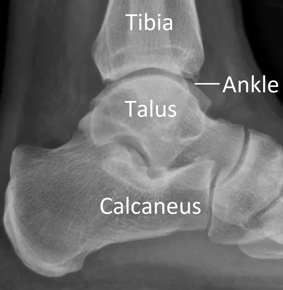
The ankle is the joint between the leg and foot. It is like a hinge but allows rotation as well.
Bones
It is composed of three bones (see images below):
- tibia
- fibula
- talus.
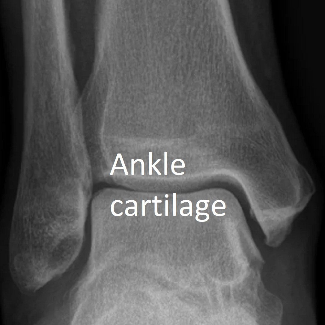
Capsule and cartilage
The joint is protected by a fibrous membrane called a joint capsule and filled with synovial fluid.
Like most joints in the human body, the ankle is covered by hyaline cartilage.
- This smooth white surface allow joint movement without friction and adds cushioning.
- There are no nerves to detect pain in cartilage but the underlying bone and adjacent tissues have many.
- Cartilage also has a poor blood supply and doesn't heal well.
- Cartilage is the "gap" between bones on X-rays (see X-ray below).

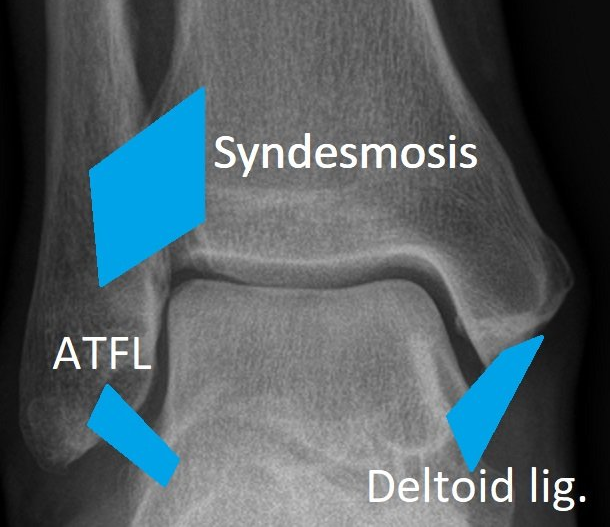
Ligaments
Ligaments are soft tissues that hold bones together (see X-ray, MRI and models below).
Ankle ligaments are divided into "high" and "low".
"High" ankle ligaments
are also known as the syndesmotic ligaments (AITFL and PITFL).
- They hold the two bones of the leg (tibia and fibula) together above the ankle.
- This connection is called the syndesmosis.
- It allows a small amount of movement between the leg bones during activities.
"Low" ankle ligaments
allow ankle movement but still hold the ankle stable.
The important ones are:
- Deltoid ligament - inside (medial) ankle.
- Anterior talo-fibular ligament (ATFL) - outside (lateral) ankle.
- Calcaneo-fibular ligament (CFL) - outside (lateral) ankle.
Tendons
Tendons connect muscles to bones and make joints move (see X-ray, MRI and models below).
The peroneal tendons
run down the outside of the leg and ankle to connect with the foot.
- Peroneus brevis.
- Peroneus longus.
- They both act to keep the ankle and foot stable on uneven ground.
There are other important tendons passing down from the leg to the foot across the ankle.
- Tibialis anterior.
- Toe flexor and extensor tendons.
- Achilles tendon.
- Tibialis posterior.